
Kliniczne aspekty chorób skóry: część 2 | Clinical Aspects of Skin Diseases: part 2
Psoriasis
Psoriasis is a chronic autoimmune skin condition characterized by the rapid turnover of skin cells, leading to the formation of red, scaly patches on the skin. It primarily affects the scalp, elbows, knees, and lower back but can appear anywhere on the body. The condition is not contagious and is often triggered by factors such as stress, infections, or skin injuries.
Pathophysiology
Psoriasis is a chronic autoimmune condition characterized by rapid turnover of epidermal keratinocytes. The pathogenesis involves:
- Immune dysregulation: Psoriasis is driven by T-cell–mediated inflammation, with Th17 and Th1 cells playing a critical role. These cells release cytokines such as IL-17 and TNF-alpha, which promote the proliferation of keratinocytes and inflammation.
- Genetic predisposition: A family history of psoriasis increases the likelihood of developing the condition, with several genetic loci (e.g., PSORS1) implicated.
- Environmental triggers: Stress, infections, medications (e.g., beta-blockers, lithium), and trauma to the skin (Koebner phenomenon) can precipitate flares.
Clinical Presentation
Psoriasis is marked by well-demarcated erythematous plaques with silvery scales, commonly found on the scalp, elbows, knees, and lower back.
Types of Psoriasis
| Type of Psoriasis | Description |
|---|---|
| Plaque Psoriasis | Thick, red plaques covered with silvery-white scales. |
| Guttate Psoriasis | Small, drop-like lesions, often triggered by streptococcal infections. |
| Inverse Psoriasis | Occurs in skin folds, with smooth, red patches. |
| Pustular Psoriasis | Characterized by white pustules surrounded by red skin. |
| Erythrodermic Psoriasis | A severe, potentially life-threatening form that involves widespread redness and scaling. |
Diagnosis
Diagnosis is clinical, based on the characteristic appearance of plaques. A biopsy may be performed to rule out other conditions in atypical cases. Key histological features include acanthosis (epidermal hyperplasia), parakeratosis (retention of nuclei in the stratum corneum), and Munro microabscesses (collections of neutrophils in the epidermis).
Treatment
Psoriasis treatment is based on the severity of the disease:
- Topical treatments: Corticosteroids, vitamin D analogs (e.g., calcipotriol), and retinoids (e.g., tazarotene) are used for mild to moderate cases.
- Phototherapy: UVB phototherapy or PUVA (psoralen + UVA) is effective for more extensive disease.
- Systemic treatments: For moderate to severe cases, methotrexate, cyclosporine, and biologics targeting TNF-alpha, IL-17, and IL-23 (e.g., adalimumab, secukinumab, ustekinumab) are used.
More About Psoriasis
- Signs: scraping a plaque produces the “candle grease” sign (silvery scale), and removing the scale reveals pinpoint bleeding (Auspitz sign). New plaques appear at sites of injury (Koebner phenomenon). Nail changes (pitting, onycholysis, oil-drop spots) are seen in about half of patients and predict psoriatic arthritis.
- Severity: measured by the percentage of body surface involved (BSA), PASI (Psoriasis Area and Severity Index) and the DLQI (quality of life). Psoriasis on the face, scalp, nails, genitals, palms and soles is more disabling than its area suggests.
- Triggers: streptococcal throat infection (guttate psoriasis), stress, obesity, smoking, alcohol, and drugs such as beta-blockers, lithium, antimalarials and NSAIDs. Systemic corticosteroids should be avoided, because stopping them can cause a severe flare of pustular or erythrodermic psoriasis.
- Red flag: generalized pustular psoriasis and erythroderma with fever, malaise and low blood pressure need urgent hospital treatment.
- Methotrexate is taken once a week, never daily (daily dosing by mistake causes fatal marrow suppression), together with folic acid; monitor blood count, liver tests and kidney function. Cyclosporine is nephrotoxic and raises blood pressure, so it is used for limited periods. Newer options include apremilast, deucravacitinib and biologics against IL-17 (secukinumab, ixekizumab) and IL-23 (guselkumab, risankizumab).
- Biologics in practice: before starting a TNF inhibitor or other biologic, exclude latent tuberculosis and hepatitis B and C. In Poland biologic treatment is available through an NFZ drug program, which depends on the severity of the disease (PASI, BSA, DLQI) and on failure or contraindication of the classic systemic drugs.
- Psoriatic arthritis: ask about morning stiffness, swollen fingers or toes (dactylitis) and heel pain. Early referral to a rheumatologist prevents joint damage.
Complications
Psoriasis is associated with several comorbidities, including psoriatic arthritis, cardiovascular disease, and metabolic syndrome. Psoriatic arthritis affects up to 30% of psoriasis patients and can lead to joint destruction if untreated.
Prognosis
- Mild Psoriasis: About 70 to 80% of patients have mild to moderate disease, which can usually be controlled with topical treatment and phototherapy. Psoriasis is nevertheless a lifelong disease with relapses and remissions, so the aim of treatment is control, not cure.
- Moderate to Severe Psoriasis: Long-term outcomes are improved with systemic therapies, but the presence of comorbidities like cardiovascular disease can complicate management. Approximately 10-30% of patients develop psoriatic arthritis.
Basal Cell Carcinoma (BCC)
Basal cell carcinoma (BCC) is a common skin cancer originating from the basal cells in the epidermis. It typically manifests as a pearly bump or a flat lesion in sun-exposed areas, such as the face and neck. BCC grows slowly and is unlikely to metastasize, making early detection and treatment important.
Pathophysiology
Basal cell carcinoma is the most common type of skin cancer, arising from the basal cells in the epidermis. It is caused by DNA mutations, primarily due to UV radiation exposure, leading to uncontrolled cell growth. BCC is a locally malignant tumor and rarely metastasizes.
Clinical Presentation
- Nodular BCC: The most common form, appearing as a pearly or translucent nodule with telangiectasia (visible blood vessels) and a rolled border. These lesions are often found on sun-exposed areas such as the face and neck.
- Superficial BCC: Appears as a scaly, red patch and is more commonly found on chest or upper back, arms, or legs.
- Morpheaform BCC: A more aggressive subtype that appears as a scar-like, ill-defined lesion.
Diagnosis
A clinical diagnosis is typically followed by biopsy to confirm the histopathology. BCC shows basaloid cells in the epidermis, with palisading nuclei and clefts between tumor cells and stroma.
Treatment
- Surgical excision: The treatment of choice for most BCCs, ensuring complete removal with clear margins.
- Mohs micrographic surgery: Often used for high-risk or facial tumors to minimize tissue loss and maximize cure rates.
- Topical therapies: For superficial BCC, imiquimod or 5-fluorouracil may be used.
- Radiation therapy: For patients who cannot undergo surgery.
- Targeted therapy: For advanced or metastatic BCC, vismodegib (a hedgehog pathway inhibitor) is an option.
Prognosis
- Excellent Prognosis: Over 95% of cases are successfully treated with surgical excision. Metastasis occurs in less than 0.5% of cases, but recurrence rates can be up to 10%, especially in high-risk areas. Regular monitoring is crucial due to the 40-50% chance of developing another BCC within five years.
Related lesions: BCC is also more likely in people with Gorlin syndrome (inherited mutations in the PTCH1 gene), and the hedgehog inhibitors vismodegib and sonidegib act on this pathway. The commonest precursor of SCC is the actinic keratosis, a rough, scaly patch on sun-damaged skin of fair-skinned older people. It is treated with cryotherapy, topical 5-fluorouracil, imiquimod, diclofenac or photodynamic therapy. Bowen disease is SCC in situ (a slowly growing red scaly plaque) and keratoacanthoma is a fast-growing dome-shaped nodule with a central keratin plug that is treated like SCC.
Squamous Cell Carcinoma (SCC)
Squamous cell carcinoma (SCC) is a type of skin cancer that originates in the squamous cells of the epidermis. It commonly appears as a firm, red nodule, a flat sore, or a scaly patch, often on sun-exposed areas such as the face, ears, and hands. SCC can grow more aggressively than basal cell carcinoma and has a higher risk of metastasis if not treated promptly.
Pathophysiology
Squamous cell carcinoma arises from keratinocytes in the epidermis and can develop from actinic keratoses, which are premalignant lesions caused by chronic UV exposure. Other risk factors include immunosuppression, HPV infection, and chronic wounds.
Clinical Presentation
SCC typically appears as a firm, red nodule or a scaly, crusted lesion. It may ulcerate or bleed, particularly in sun-exposed areas such as the face, neck, arms, and hands. High-risk SCCs include lesions on the ears, lips, and genitalia, as well as those that are large, deep, or rapidly growing.
Diagnosis
Diagnosis is confirmed by biopsy, revealing atypical keratinocytes extending beyond the epidermis into the dermis. Keratin pearls are often seen histologically.
Treatment
- Surgical excision with clear margins is the primary treatment.
- Mohs micrographic surgery is preferred for high-risk lesions or those located on the face, hands, or feet.
- Cryotherapy or electrodessication and curettage (ED&C) may be used for superficial SCCs.
- Radiation therapy is an option for patients who are not surgical candidates.
- For advanced or metastatic SCC, systemic chemotherapy or immunotherapy (e.g., cemiplimab, an anti-PD-1 inhibitor) may be used.
Prognosis
- Favorable with Early Detection: When treated early, the cure rate exceeds 90%. However, high-risk lesions have a 2-5% chance of metastasis. Advanced SCC has a more guarded prognosis, and the 5-year survival falls substantially once lymph nodes or distant organs are involved.
Melanoma
Melanoma is a type of skin cancer that develops in melanocytes, the cells responsible for producing melanin, the pigment that gives skin its color. While it primarily occurs in the skin, it can also manifest in other parts of the body, such as the eyes, mucous membranes, and rarely in internal organs.
Melanoma has seen a steady increase in incidence over the past few decades, especially in regions with high sun exposure. Risk factors include:
- UV Radiation: Overexposure to ultraviolet (UV) radiation, either from the sun or artificial sources like tanning beds.
- Fair Skin: People with fair skin, light-colored eyes, and a tendency to burn rather than tan are at a higher risk.
- Family History: Individuals with a family history of melanoma have an increased risk.
- Multiple Moles: Presence of numerous moles on the body can elevate the risk of developing melanoma.
- Immune Suppression: People with weakened immune systems due to certain medical conditions or medications have an increased susceptibility.
Diagnosis of Melanoma
A dermatologist typically performs a thorough skin examination to evaluate moles for suspicious characteristics. Two widely used systems aid in evaluating suspicious skin lesions: the ABCDE criteria and the Revised 7-Point Checklist. However, a biopsy of the suspicious mole or skin lesion is the definitive method to confirm melanoma.
ABCDE Criteria
This system is commonly employed to identify suspicious moles that may indicate melanoma:
- A – Asymmetry: One half of the lesion differs from the other.
- B – Border: The borders are irregular, notched, or poorly defined.
- C – Color: Variations in color, including different shades of brown, black, or even red, white, or blue.
- D – Diameter: Moles larger than 6 mm (about the size of a pencil eraser) should be examined more closely.
- E – Evolution: Changes in size, shape, color, or symptoms such as itching or bleeding are concerning and should be evaluated promptly.
Revised 7-Point Checklist
This method provides a more structured approach for assessing lesions. It assigns points to major and minor criteria:
- Major Criteria (2 points each): Change in size, shape, or color.
- Minor Criteria (1 point each): Inflammation, crusting or bleeding, sensory changes, and diameter ≥7 mm.
Any patient scoring three or more points on the checklist should be referred for further dermatological evaluation.
Stages of melanoma
Melanoma is divided into stages using five Roman numerals (0 through IV) and up to four letters (A through D) that indicate a higher risk within each stage. Cancer treatment options and prognoses are dependent on the stage of cancer.
The stages of melanoma are determined mostly by specific details about the tumor and its growth that are tallied in a system called TNM where:
T stands for Tumor,
N stands for Nodes,
M stands for Metastasis.
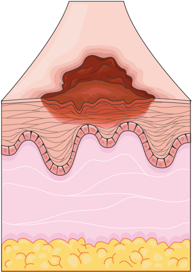
Stage 0 (In Situ): Melanoma is confined to the epidermis, with no invasion of deeper layers.
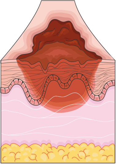
Stage I: Localized melanoma that is still thin (up to 1 mm, or up to 2 mm if it is not ulcerated), confined to the skin.
Stage II: Thicker melanoma (over 2 mm, or over 1 mm if it is ulcerated) but still localized to the skin, with no lymph node involvement.
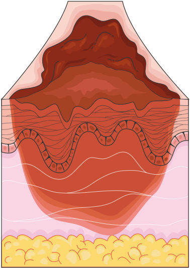
Stage III: Melanoma has spread to nearby lymph nodes or tissues but has not yet reached distant organs.
Stage IV: Advanced melanoma with distant metastases, affecting organs such as the lungs, liver, brain, or bones.
Treatment Options
The treatment of melanoma depends on the stage of the disease:
- Surgical Excision: The primary treatment for localized melanoma involves excising the lesion along with a margin of healthy skin to ensure complete removal.
- Immunotherapy: Drugs like checkpoint inhibitors (e.g., nivolumab, pembrolizumab) are used to enhance the body’s immune response to melanoma cells.
- Targeted Therapy: Used for melanomas with specific genetic mutations (e.g., BRAF mutations), BRAF inhibitors (vemurafenib, dabrafenib, encorafenib) are combined with MEK inhibitors (cobimetinib, trametinib, binimetinib) to inhibit cancer cell growth.
- Chemotherapy: While less commonly used for melanoma today, chemotherapy may still be considered in advanced stages or if other treatments are ineffective.
- Radiation Therapy: This may be employed to treat areas of melanoma that have spread, particularly in palliative care settings.
Prevention Strategies
Preventing melanoma revolves around minimizing UV radiation exposure:
- Regular Skin Checks: Patients, especially those with fair skin, a history of sunburns, or a family history of melanoma, should undergo regular skin examinations and self-checks for new or changing moles.
- Sunscreen: Regular use of broad-spectrum sunscreen with an SPF of 30 or higher is vital for protection.
- Protective Clothing: Wearing hats, sunglasses, and long sleeves when outdoors provides a physical barrier against UV rays.
- Avoid Tanning Beds: Artificial UV sources increase the risk of melanoma and should be avoided.
Prognosis
- Early-Stage Melanoma: Stage 0-I melanoma has a 5-year survival rate of over 90-99% with surgical excision.
- Advanced Melanoma: Survival rates decrease significantly: in stage III the 5-year survival ranges from about 30% to over 90% depending on the substage, and stage IV used to have a 5-year survival of only 15 to 20%. Advances in treatment, like immunotherapy, have improved survival rates in recent years.
Melanoma Subtypes, Surgical Margins and Sentinel Node Biopsy
| Subtype | Features |
|---|---|
| Superficial spreading | The most common (about 70%); a flat, irregular, multicolored patch that slowly enlarges |
| Nodular | Fast-growing, often dark-red or black, symmetrical dome; may be amelanotic; invades deeply early |
| Lentigo maligna melanoma | Older adults, chronically sun-damaged face; develops from lentigo maligna (melanoma in situ) |
| Acral lentiginous | Palms, soles and under the nail; the main type in people with dark skin; often diagnosed late |
The most important prognostic factor is the Breslow thickness, which is the depth of the tumor in millimeters, measured from the granular layer to the deepest tumor cell. After the diagnostic excision biopsy, wide local excision is performed with margins that depend on the thickness: 0.5 to 1 cm for melanoma in situ, 1 cm for tumors up to 1 mm, 1 to 2 cm for tumors of 1 to 2 mm, and 2 cm for thicker tumors. A sentinel lymph node biopsy is offered for tumors thicker than about 0.8 mm (or thinner but ulcerated) to stage the disease. Patients with stage III and IV melanoma are treated with immune checkpoint inhibitors (anti-PD-1, sometimes with anti-CTLA-4) or, if there is a BRAF V600 mutation, with BRAF and MEK inhibitors. Adjuvant immunotherapy after surgery is used in stage III and in high-risk stage II disease.
Merkel Cell Carcinoma (MCC)
Merkel cell carcinoma is a rare but highly aggressive neuroendocrine skin cancer, arising from Merkel cells in the epidermis. Although far less common than basal cell carcinoma, squamous cell carcinoma, or melanoma, it carries a notably poorer prognosis, with the 5-year survival falling from about three quarters in localized disease to around one half or less once regional lymph nodes are involved, and to roughly a quarter or less with distant metastasis.
Risk factors include advanced age, chronic UV exposure, fair skin, and immune suppression (e.g., organ transplant recipients, HIV). Most cases are linked to the Merkel cell polyomavirus.
It typically presents as a rapidly growing, painless, firm, red or violaceous nodule on sun-exposed skin, often remembered by the mnemonic AEIOU: Asymptomatic, Expanding rapidly, Immune suppression, Older than 50, and Ultraviolet-exposed site.
Diagnosis requires biopsy with immunohistochemical staining. Treatment involves wide surgical excision, often combined with sentinel lymph node biopsy and adjuvant radiation, given the high rate of early lymphatic spread. Immunotherapy (e.g., avelumab) is used for advanced or metastatic disease.
Other Skin Conditions to Recognize
- Erythema multiforme: an acute reaction, most often to herpes simplex virus or Mycoplasma infection, with symmetrical target lesions (three zones: a dark center, a pale ring and a red outer ring), mainly on the hands, feet and limbs. It is self-limiting. Do not confuse it with Stevens-Johnson syndrome and toxic epidermal necrolysis, which are usually drug-induced, with widespread blistering, mucosal erosions, and skin detachment.
- Hidradenitis suppurativa: a chronic inflammatory disease of the skin folds (axillae, groin, under the breasts) with painful nodules, abscesses, draining sinus tracts and scarring. Smoking and obesity are risk factors. Treatment depends on severity (Hurley stages): antiseptic washes and topical clindamycin, oral tetracyclines, adalimumab or secukinumab, and surgical excision for severe disease.
- Dermatofibroma: a firm, benign, brown nodule, usually on the legs, which dimples inwards when pinched (the dimple sign). Reassurance is enough.
- Pityriasis rosea: a self-limiting rash in young adults that starts with a single oval “herald patch” on the trunk, followed after one to two weeks by smaller scaly patches in a “Christmas tree” pattern along the skin lines. It resolves in 6 to 8 weeks; check the syphilis serology if the picture is atypical.
- Angioedema: swelling of the deep dermis and subcutaneous tissue of the lips, eyelids, tongue and larynx. Histamine-mediated angioedema (allergy, with urticaria) responds to epinephrine (adrenaline) and antihistamines. Bradykinin-mediated angioedema (ACE inhibitors, or hereditary C1-esterase inhibitor deficiency) occurs without urticaria and does not respond to these drugs; it is treated with C1-inhibitor concentrate or icatibant. Airway swelling is an emergency.
- Frostbite: freezing injury of the fingers, toes, nose and ears. The skin is white and numb, then red, blistered, and finally black if it dies. Rewarm in water at 37 to 39 °C only when there is no risk of refreezing, never rub the skin or use dry heat, give ibuprofen for pain, and treat severe cases with iloprost or thrombolysis. A person should not walk on frozen feet before rewarming.
Rashes in Childhood Infections
- Measles: fever, cough, coryza and conjunctivitis, Koplik spots on the buccal mucosa, and then a red maculopapular rash that spreads from the face downward. Complications are otitis media, pneumonia, encephalitis and subacute sclerosing panencephalitis. It is prevented by MMR vaccination.
- Rubella: a mild illness with a rash and tender lymph nodes behind the ears. Infection in early pregnancy causes congenital rubella syndrome (cataract, deafness, heart defects).
- Scarlet fever: caused by group A streptococcus, with a fine sandpaper-like rash, a strawberry tongue, a flushed face with perioral pallor and a sore throat. Treat with phenoxymethylpenicillin for 10 days. Late complications are rheumatic fever and glomerulonephritis.
- Hand, foot and mouth disease: caused by coxsackievirus, with painful mouth ulcers and vesicles on the palms and soles in young children. It is self-limiting.
- Erythema infectiosum (fifth disease): parvovirus B19, with “slapped cheek” redness followed by a lacy rash. It can cause a transient aplastic crisis in sickle cell disease and hydrops fetalis in pregnancy.
- Roseola infantum: human herpesvirus 6; a high fever for three days in a toddler, followed by a rash as the temperature falls. Febrile seizures can occur.
Bacterial Skin and Soft Tissue Infections
Skin infections are among the most common reasons for a visit to a general practitioner or an emergency department. The most frequent bacteria are Staphylococcus aureus and Streptococcus pyogenes (group A streptococcus). The depth of the infection decides how serious it is, from the epidermis (impetigo) through the dermis and subcutaneous tissue (erysipelas and cellulitis) to the fascia (necrotizing fasciitis).
Impetigo
Impetigo is a superficial, highly contagious infection of the epidermis, most common in young children. The non-bullous form starts as small vesicles that rupture and leave honey-colored crusts, usually around the nose and mouth. The bullous form, caused by toxin-producing S. aureus, produces flaccid blisters. Localized impetigo is treated with a topical antibiotic (mupirocin or fusidic acid) for about 5 to 7 days. Extensive or bullous disease needs an oral antibiotic active against staphylococci and streptococci, such as flucloxacillin or cefalexin (in Poland, cefalexin or cefadroxil are usual). The child should stay away from school until the lesions have crusted over or until 48 hours of treatment have passed. A late complication of streptococcal impetigo is post-streptococcal glomerulonephritis.
Erysipelas and Cellulitis
Erysipelas is an infection of the upper dermis, mostly caused by streptococci. It appears as a bright red, raised, sharply demarcated plaque, most often on the face or the lower leg, with fever and chills. Cellulitis involves the deeper dermis and the subcutaneous tissue, and its borders are less well defined. Both present with erythema, warmth, swelling and tenderness. Risk factors are a skin break (tinea pedis, an insect bite, a wound), lymphedema, venous insufficiency, obesity and diabetes.
- Diagnosis: clinical. Mark the edge of the redness with a pen to see if it spreads. Blood tests (CRP, white cell count) and blood cultures are used in patients with fever or systemic signs. Remember the mimics: deep vein thrombosis, stasis dermatitis, gout and contact dermatitis. Cellulitis is usually unilateral, whereas stasis dermatitis tends to be bilateral.
- Treatment: mild disease is treated at home with an oral penicillin (phenoxymethylpenicillin or amoxicillin) or, if staphylococci are suspected, with flucloxacillin or a cephalosporin, usually for 5 to 7 days. Patients with fever, rapid spread, hypotension, a very young or old age or poor response need admission and intravenous antibiotics (for example cefazolin or ceftriaxone; vancomycin if MRSA is a risk). Elevate the limb and treat the cause, such as athlete’s foot or edema.
- Complications: abscess formation, bacteremia and sepsis, and recurrence. After two or more episodes a year, long-term prophylaxis with penicillin is considered.
Cutaneous Abscess and Furuncle
A furuncle (boil) is an infection of a hair follicle, and a carbuncle is a cluster of connected furuncles. A cutaneous abscess is a painful, red, fluctuant collection of pus. The treatment of choice is incision and drainage. Antibiotics are added only when there is surrounding cellulitis, fever, a lesion on the face or in an immunosuppressed or diabetic patient, or when there are multiple abscesses. Recurrent abscesses suggest MRSA carriage, and nasal and skin decolonization is considered.
Necrotizing Fasciitis
Necrotizing fasciitis is a rare, life-threatening infection of the fascia and subcutaneous tissue, often polymicrobial or caused by group A streptococcus. The key warning sign is pain out of proportion to the physical findings, followed by rapidly spreading redness, swelling, blisters, dusky or black skin, crepitus and septic shock. Imaging must not delay treatment. The treatment is urgent surgical debridement and broad-spectrum intravenous antibiotics (for example piperacillin-tazobactam or a carbapenem, with vancomycin and clindamycin). Mortality remains high, particularly when treatment is delayed.
Viral Skin Infections
Herpes Simplex
Herpes simplex virus type 1 usually causes cold sores (herpes labialis) and type 2 usually causes genital herpes, although either can infect either site. After the primary infection the virus stays latent in a sensory ganglion and reactivates with stress, fever, sun or illness. Typical lesions are grouped vesicles on a red base that crust over within a week. A tingling or burning sensation (prodrome) often comes first. Treatment of frequent or severe episodes is oral acyclovir (for example 200 mg five times a day for 5 days) or valacyclovir. Treatment is most effective if started early. Dangerous forms are eczema herpeticum (see atopic dermatitis), herpes keratitis, which threatens the eye, herpes in the newborn and widespread infection in immunosuppressed patients, who may need intravenous acyclovir.
Herpes Zoster (Shingles)
Shingles is the reactivation of the varicella-zoster virus that has remained latent in the dorsal root ganglia after chickenpox. It is more frequent in older adults and in immunosuppressed patients. A burning, stabbing or tingling pain in one dermatome precedes the rash by 1 to 3 days. Then grouped vesicles appear on a red base in a unilateral, band-like distribution that does not cross the midline. The lesions crust over in 7 to 10 days.
- Treatment: antiviral treatment (acyclovir 800 mg five times a day, valacyclovir 1 g three times a day or famciclovir 500 mg three times a day, for 7 days) should be started within 72 hours of the rash, and later if new vesicles are still appearing or if the patient is over 50, immunosuppressed or has a complicated form. Analgesia is needed, often with a gabapentinoid or a tricyclic antidepressant for neuropathic pain.
- Urgent forms: herpes zoster ophthalmicus (the first branch of the trigeminal nerve; vesicles on the tip of the nose, called the Hutchinson sign, suggest eye involvement) needs same-day ophthalmological assessment. Ramsay Hunt syndrome (vesicles in the ear canal, ear pain, facial palsy) needs urgent treatment with antivirals and a corticosteroid. Disseminated zoster in immunosuppressed patients needs intravenous acyclovir.
- Complications: postherpetic neuralgia (pain that persists for more than 3 months and is more common in older patients), scarring and secondary infection.
- Prevention: the recombinant adjuvanted zoster vaccine (two doses) is recommended for adults aged 50 and over and for immunocompromised adults.
Warts and Molluscum Contagiosum
Common warts (verrucae) are caused by human papillomavirus (HPV), mostly types 1, 2 and 4. They are rough, hyperkeratotic papules on the hands and feet, and plantar warts are painful when walking. Many warts disappear spontaneously within two years, especially in children. Treatment is salicylic acid, cryotherapy or watchful waiting. Genital warts (condylomata acuminata) are caused by HPV types 6 and 11 and are sexually transmitted. Molluscum contagiosum is caused by a poxvirus and gives pearly, dome-shaped papules with a central dimple (umbilication), mostly in children, and in adults when it is sexually acquired. It resolves by itself in 6 to 18 months in healthy people.
Fungal Skin Infections
Superficial fungal infections are very common. Dermatophytes (Trichophyton, Microsporum, Epidermophyton) feed on keratin and cause tinea, also called ringworm. The site gives the name: tinea corporis (body), tinea cruris (groin), tinea pedis (feet), tinea capitis (scalp) and tinea unguium or onychomycosis (nails).
- Tinea corporis: a ring-shaped (annular) red plaque with an active, scaly edge and central clearing. Itchy.
- Tinea pedis (athlete’s foot): scaling, maceration and fissures between the toes, or a dry “moccasin” scaling of the sole. It is a common entry point for cellulitis.
- Tinea cruris: a red, sharply demarcated rash in the groin that usually spares the scrotum (unlike candidiasis).
- Tinea capitis: mostly in children. Scaly patches with hair loss or broken hairs, and sometimes a boggy, inflamed swelling called kerion. It needs oral treatment.
- Onychomycosis: thickened, yellow, crumbling nails, mostly the toenails.
- Pityriasis versicolor: caused by the yeast Malassezia. Fine scaly patches, lighter or darker than the surrounding skin, on the trunk and shoulders in warm weather. Treated with ketoconazole or selenium sulfide shampoo.
- Cutaneous candidiasis: a bright red, moist rash with small satellite lesions in the skin folds (intertrigo) and in the diaper area of babies. It is more frequent in diabetes, obesity and after antibiotics.
Diagnosis: a skin scraping, a nail clipping or hair is examined directly with potassium hydroxide (KOH) and sent for fungal culture. Nail disease must be confirmed in the laboratory before long oral treatment. Treatment: topical terbinafine or an azole (clotrimazole, miconazole) for 2 to 4 weeks. Oral terbinafine is used for extensive disease, for tinea capitis (griseofulvin is preferred for Microsporum) and for onychomycosis (250 mg daily for about 6 weeks for fingernails and 12 weeks for toenails, with liver tests). Topical corticosteroids must not be used alone, because they make the rash less typical (tinea incognito) and worse.
Scabies and Head Lice
Scabies
Scabies is an infestation with the mite Sarcoptes scabiei. It spreads by prolonged skin contact and within families and care homes. The cardinal symptom is intense itching that is worse at night, starting 3 to 6 weeks after the first infestation (earlier on re-exposure). The mite digs thin, wavy, gray burrows in the finger webs, wrists, elbows, axillae, waistline and genitals, and small itchy papules and excoriations appear. The head and neck are spared in adults but are involved in infants. Crusted (Norwegian) scabies occurs in immunosuppressed and elderly patients and is extremely contagious.
- Diagnosis: clinical, with dermoscopy or microscopy of a skin scraping to find mites, eggs or feces.
- Treatment: permethrin 5% cream applied from the neck to the toes (including the head in infants and in the elderly), left for 8 to 12 hours, and repeated after 7 days. Oral ivermectin (200 micrograms/kg, repeated after 7 to 14 days) is an alternative, especially in outbreaks and in crusted scabies. At the same time all household members and close contacts are treated, even without symptoms. Bedding, towels and clothes are washed at 60 °C or sealed in a bag for 3 days. The itch can continue for 2 to 4 weeks after successful treatment (post-scabetic itch).
Head Lice (Pediculosis Capitis)
Head lice are common in school children. They cause itching of the scalp, especially behind the ears and at the nape, and the diagnosis is made by finding live lice, because empty nits alone do not prove active infestation. Treatment is a dimeticone lotion or permethrin 1%, combined with wet combing, and repeated after 7 to 10 days. Close contacts are examined.
Urticaria
Urticaria (hives) is characterized by wheals: itchy, raised, pink or red swellings that last less than 24 hours and move around, leaving normal skin. It is often accompanied by angioedema. Acute urticaria lasts less than 6 weeks and is usually caused by a viral infection, a drug (antibiotics, NSAIDs, aspirin) or food. Chronic urticaria lasts more than 6 weeks. Most cases are chronic spontaneous urticaria, which is autoimmune in many patients, and some are inducible (cold, pressure, heat, exercise, sunlight, dermographism). The cause is rarely found, so extensive tests are not needed.
- Diagnosis: clinical. In chronic disease, tests are limited to a blood count, CRP and sometimes thyroid function, unless the history suggests a cause. Wheals that last more than 24 hours or leave bruising suggest urticarial vasculitis, and a skin biopsy is needed.
- Treatment: avoid triggers and NSAIDs. The first choice is a non-sedating second-generation antihistamine (cetirizine, loratadine, bilastine). If it does not help, the dose can be increased up to four times the standard dose. Omalizumab is used in patients who do not respond, and short courses of oral corticosteroids for severe acute episodes only.
- Emergency: hives with swelling of the tongue or throat, wheeze or hypotension is anaphylaxis and is treated with intramuscular epinephrine.
Rosacea and Seborrheic Dermatitis
Rosacea
Rosacea is a chronic inflammatory disease of the central face, mostly in fair-skinned adults over 30. It shows as persistent redness, flushing, visible small vessels (telangiectasia) and, in the papulopustular type, papules and pustules without comedones, which distinguishes it from acne. Triggers are sun, heat, alcohol, hot drinks and spicy food. In some men a thickened, bulbous nose (rhinophyma) develops, and eye involvement (ocular rosacea) causes a gritty, red eye. Treatment is sun protection and avoidance of triggers, topical metronidazole, ivermectin or azelaic acid, and in more severe disease low-dose oral doxycycline. Topical steroids on the face should be avoided because they worsen rosacea.
Seborrheic Dermatitis
Seborrheic dermatitis is a common, chronic, relapsing condition involving the sebum-rich areas: the scalp (dandruff), the eyebrows, the nasolabial folds, behind the ears and the chest. The skin is red with greasy, yellowish scales. The yeast Malassezia and an inflammatory reaction play a role. In infants it appears as cradle cap, which is harmless and resolves by itself. Severe or sudden disease can accompany HIV infection and Parkinson disease. Treatment is an antifungal shampoo or cream (ketoconazole 2%), and a short course of a mild topical corticosteroid or a calcineurin inhibitor for flares.
Vitiligo and Hair Loss
Vitiligo
Vitiligo is an acquired autoimmune loss of melanocytes. It gives well-demarcated, milk-white (depigmented) patches, typically around the mouth and eyes, on the hands, elbows, knees and genitals, often symmetrical. It affects about 1% of people. It is associated with other autoimmune diseases, especially autoimmune thyroid disease, type 1 diabetes, pernicious anemia and Addison disease, so thyroid function should be checked. A Wood’s lamp shows the patches clearly. Treatment aims to stop progression and to repopulate the skin: topical corticosteroids or calcineurin inhibitors, narrow-band UVB phototherapy and, more recently, topical ruxolitinib (a JAK inhibitor). Sun protection is important, because the depigmented skin burns easily. The condition has a large psychological impact.
Hair Loss: the Common Types
- Androgenetic alopecia: the most common cause. In men it starts with temporal recession and a thinning crown, and in women it shows as a widening parting with the hairline preserved. Treatment: topical minoxidil, and finasteride in men.
- Telogen effluvium: diffuse shedding 2 to 3 months after a trigger such as childbirth, high fever, major surgery, severe stress, crash dieting, iron or thyroid disease, or a new drug. It is reversible in most patients once the trigger is removed. The hair pull test is positive.
- Alopecia areata: an autoimmune disease with round, smooth patches of non-scarring hair loss, and “exclamation mark” hairs at the edge. It can progress to total scalp (alopecia totalis) or body (alopecia universalis) loss. Treatment: topical or intralesional corticosteroids, and JAK inhibitors (baricitinib, ritlecitinib) for severe disease. It is linked with other autoimmune diseases and with atopy.
- Scarring alopecia (for example discoid lupus or lichen planopilaris): the follicles are destroyed, the skin is smooth and shiny and without follicular openings, and the hair does not regrow. Early referral and biopsy are necessary.
- Tinea capitis: a fungal cause that must not be missed in children (see above).
Chronic Wounds: Pressure Ulcers and Leg Ulcers
Pressure Ulcers (Pressure Injuries)
A pressure ulcer is damage to the skin and the tissue under it caused by prolonged pressure, or pressure with shear. It develops over bony prominences: the sacrum, heels, hips (greater trochanters), ischial tuberosities and the back of the head. The main risk factors are immobility, incontinence and moist skin, poor nutrition, sensory loss (for example after a stroke or spinal cord injury), older age and critical illness. Risk is assessed with a scale such as the Braden scale.
| Stage | Findings |
|---|---|
| Stage 1 | Intact skin with non-blanchable erythema |
| Stage 2 | Partial-thickness skin loss: a shallow open ulcer with a pink wound bed, or an intact or ruptured blister |
| Stage 3 | Full-thickness skin loss; subcutaneous fat is visible, but not fascia, muscle or bone |
| Stage 4 | Full-thickness loss with exposed fascia, muscle, tendon, ligament, cartilage or bone |
| Unstageable | The wound bed is covered by slough or eschar, so the depth cannot be seen |
| Deep tissue injury | Persistent deep red, maroon or purple discoloration of intact skin |
- Prevention: repositioning at regular intervals (usually every 2 to 4 hours, individualized), pressure-redistributing mattresses and cushions, keeping the heels free of pressure, keeping the skin clean and dry, managing incontinence, and adequate nutrition and protein intake.
- Treatment: remove pressure from the area, clean the wound, remove dead tissue (debridement), choose a moist dressing, treat infection and optimize nutrition. Suspect osteomyelitis in a deep ulcer over bone. Surgery is considered for stage 3 and 4 ulcers.
Leg Ulcers
Most leg ulcers are venous, arterial or neuropathic (diabetic), and often there is more than one cause. Distinguishing between them is a key clinical skill, because the treatments are different, and compression therapy can be dangerous in arterial disease.
| Feature | Venous ulcer | Arterial ulcer | Diabetic neuropathic ulcer |
|---|---|---|---|
| Location | Medial malleolus and the lower third of the leg (the “gaiter” area) | Toes, heels, lateral malleolus and pressure points | Plantar surface, over metatarsal heads and heels |
| Appearance | Shallow, irregular, with moist yellow slough and exudate | Punched out, deep, dry, pale or necrotic base | Deep, with a callus around it |
| Pain | Mild to moderate, relieved by elevation | Severe, worse at night and on elevation, relieved by hanging the leg down | Painless because of neuropathy |
| Surrounding skin | Varicose veins, edema, brown hemosiderin staining, lipodermatosclerosis, venous eczema | Cool, shiny, hairless skin; weak or absent pulses; delayed capillary refill | Dry skin, loss of sensation, foot deformity |
| Key tests and treatment | Doppler study (ankle-brachial pressure index of at least 0.8) and then compression bandaging; treat the varicose veins | Ankle-brachial index below 0.8; urgent vascular referral for revascularization | Offloading, debridement, infection control and glucose control; probe-to-bone test for osteomyelitis |
Severe Cutaneous Drug Reactions
Most drug rashes are mild (a maculopapular, morbilliform eruption 4 to 14 days after the start of a drug), but a few are life-threatening and must be recognized at once. The warning signs are fever, facial swelling, painful skin, blisters, mucosal erosions, skin detachment and abnormal blood tests.
- Stevens-Johnson syndrome (SJS) and toxic epidermal necrolysis (TEN): a spectrum of the same disease, classified by the percentage of body surface area with epidermal detachment: SJS less than 10%, SJS/TEN overlap 10 to 30%, TEN more than 30%. A flu-like prodrome is followed by painful, dusky red or purpuric macules, blisters, erosions of the mouth, eyes and genitals, and a positive Nikolsky sign. It usually starts 1 to 3 weeks (up to 8 weeks) after a new drug. Typical causes are allopurinol, carbamazepine, lamotrigine, phenytoin, sulfonamides, nevirapine and NSAIDs of the oxicam type. Treatment: stop the suspected drug at once, treat in an intensive care or burns unit, give fluids, wound and eye care, and consider cyclosporine or intravenous immunoglobulin. Mortality is about 10% in SJS and 30% or more in TEN. The patient must be told to avoid the drug for life.
- DRESS (drug reaction with eosinophilia and systemic symptoms): appears 2 to 8 weeks after starting a drug (allopurinol, anticonvulsants, vancomycin, sulfonamides). Fever, a widespread rash, facial edema, lymphadenopathy, eosinophilia and organ involvement (most often the liver, also the kidneys and lungs). Treatment: stop the drug, give systemic corticosteroids, and monitor the organs.
- Fixed drug eruption: one or a few round, dusky red or purple patches that return at the same site each time the drug is taken (for example NSAIDs, acetaminophen (paracetamol) and tetracyclines), and leave pigmentation.
- Anaphylaxis and angioedema: see urticaria above.
Autoimmune Blistering Diseases
Autoimmune blistering diseases are caused by antibodies against the structures that hold the epidermis together or attach it to the dermis. The level of the split in the skin explains the type of the blister and the clinical picture (see the basement membrane zone in the anatomy lesson).
| Feature | Pemphigus vulgaris | Bullous pemphigoid | Dermatitis herpetiformis |
|---|---|---|---|
| Typical patient | Age 40 to 60 | Older than 70 | Young and middle-aged adults, often with celiac disease |
| Target antigen | Desmoglein 3 (and 1) in the desmosomes | BP180 and BP230 in the hemidesmosomes | Epidermal and tissue transglutaminase |
| Blisters | Flaccid, easily rupture, leave painful erosions; mouth involved early | Tense, large, on red or urticarial skin; flexures, limbs; mouth rarely involved | Very itchy, grouped small vesicles on the elbows, knees, buttocks |
| Nikolsky sign | Positive | Negative | Negative |
| Direct immunofluorescence | IgG between keratinocytes (chicken wire) | Linear IgG and C3 along the basement membrane | Granular IgA in the dermal papillae |
| Treatment | Oral prednisolone and rituximab; immunosuppressants | Potent topical corticosteroid (clobetasol) or oral prednisolone; doxycycline | Gluten-free diet and dapsone |
Pemphigus vulgaris is life-threatening without treatment, because of widespread erosions, infection and fluid loss. Bullous pemphigoid is more common, and in many patients it is linked to neurological disease (dementia, stroke) and to some drugs (gliptins, furosemide). Treatment of all these diseases is directed by a dermatologist.
Skin Signs of Connective Tissue Disease
- Systemic lupus erythematosus: the malar (butterfly) rash is a red rash over the cheeks and nose bridge that spares the nasolabial folds. Other signs are photosensitivity, oral ulcers, and diffuse hair loss. Discoid lupus gives well-defined, scaly red plaques with follicular plugging that heal with scarring, atrophy, pigment change and scarring alopecia. It is treated with sun protection, topical steroids and hydroxychloroquine, and a small percentage of patients develop systemic lupus.
- Dermatomyositis: a violaceous (heliotrope) rash on the upper eyelids, flat-topped violaceous papules over the knuckles (Gottron papules), a “shawl” sign of erythema over the shoulders and upper back, and dilated capillaries at the nail folds, together with symmetrical proximal muscle weakness. Check creatine kinase and myositis antibodies. In adults dermatomyositis can be a paraneoplastic syndrome, so a search for an underlying cancer is needed.
- Systemic sclerosis (scleroderma): thickening and tightening of the skin of the fingers (sclerodactyly), beak-like nose and small mouth, telangiectasia, calcinosis, and digital ulcers, nearly always preceded by Raynaud phenomenon. Morphea is a localized form limited to the skin.
Common Benign Skin Lesions
- Seborrheic keratosis: a very common, waxy, brown, “stuck-on” plaque in older adults on the trunk and face. It is harmless and needs treatment only for cosmetic reasons or irritation. The sudden appearance of many lesions at once (the Leser-Trelat sign) can accompany an internal cancer, but is rare.
- Actinic keratosis: a rough, scaly patch on sun-damaged skin that can progress to squamous cell carcinoma (see above).
- Cherry angioma: a bright red, small, dome-shaped papule made of capillaries. It increases with age and is harmless.
- Skin tags (acrochordons): soft, small pedunculated lesions in the folds, linked with obesity and diabetes.
- Lipoma: a soft, mobile, painless subcutaneous lump that grows slowly. A lump that is fast-growing, larger than 5 cm, deep or painful should be investigated for liposarcoma.
- Epidermoid cyst: a firm, round, mobile nodule with a central punctum, which can become inflamed and infected. Treatment is complete surgical excision of the cyst wall.
- Infantile hemangioma: a red “strawberry” vascular tumor that appears in the first weeks of life, grows for several months and then involutes by itself. Propranolol is used for hemangiomas that threaten vision, the airway or function, or that ulcerate.
- Hyperhidrosis: excessive sweating of the palms, axillae or soles. Treatment starts with aluminum chloride antiperspirants and goes on to iontophoresis, botulinum toxin injections and oral anticholinergics. Secondary causes include hyperthyroidism, infection and drugs.
- Acanthosis nigricans: velvety, dark thickening of the skin in the neck and axillae. It is a marker of insulin resistance and obesity, and rarely of cancer.
- Xanthelasma: yellowish plaques on the eyelids, which may be linked to high cholesterol.