Anatomia układu kostnego | Skeletal System Anatomy
Did you know?
Did you know that bones can conduct electrical currents? Research has shown that bone tissue has piezoelectric properties, meaning it can generate an electric charge in response to mechanical stress. This phenomenon is utilized in medical treatments like bone growth stimulation, where low electrical currents are applied to help heal fractures and stimulate bone regeneration. This ability of bone to convert mechanical stress into electrical energy plays a role in maintaining bone health and adapting to physical activity.
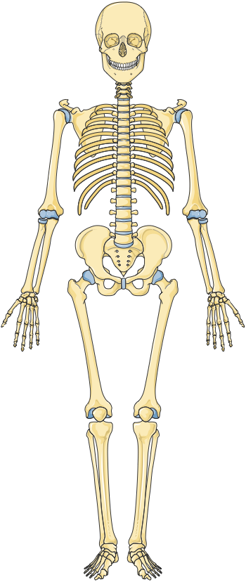

The skeletal system consists of 206 bones in adults, varying in size, shape, and function. These bones are the foundation of the human body, supporting overall health and bodily functions.
Bone Matrix
The bone matrix is the extracellular material that forms the bulk of bone tissue, consisting of both organic and inorganic components:
Organic Matrix
- Collagen Fibers: Collagen is the primary organic component of bone, accounting for about 30% of bone mass. These fibers provide flexibility and tensile strength, enabling bones to withstand forces such as stretching and twisting. Collagen also contributes to the bone’s resilience, making it less prone to fracture under stress.
- Proteoglycans and Glycoproteins: These molecules are embedded in the collagen matrix and help bind water to the bone, contributing to its resistance to compression.
Inorganic Matrix
- Mineral Salts: The predominant mineral in bone is hydroxyapatite, a crystalline structure composed mainly of calcium phosphate. This mineral component makes up about 70% of bone mass, providing hardness and rigidity, which allows bones to support body weight and resist compressive forces.
- Other Minerals: Additional minerals such as calcium carbonate, magnesium, sodium, and fluoride are present and contribute to bone strength and metabolic functions.
Bone Cells
Bone tissue is dynamic, involving various cell types responsible for bone formation, maintenance, and resorption:
| Cell Type | Description and Function |
|---|---|
| Osteoblasts | Bone-forming cells that are responsible for synthesizing and secreting the organic components of the bone matrix, including collagen and osteoid. They are found on the surface of bone tissue and play a crucial role in bone growth, mineralization, and repair. Once they have completed forming new bone, they may differentiate into osteocytes or become lining cells on the bone surface. |
| Osteocytes | Mature bone cells that originate from osteoblasts and are embedded within lacunae in the bone matrix. They have long cytoplasmic extensions that connect to other osteocytes and bone cells through tiny channels called canaliculi. Osteocytes are essential for maintaining the bone matrix, regulating mineral content, and coordinating bone remodeling. They can also detect and respond to mechanical stress, signaling osteoblasts and osteoclasts to strengthen or resorb bone as needed. |
| Osteoclasts | Large, multinucleated cells that break down (resorb) bone tissue by secreting enzymes and acids that dissolve the bone matrix, releasing calcium and other minerals into the bloodstream. Osteoclasts are crucial for bone remodeling, reshaping bone during growth, and maintaining calcium homeostasis in the body. They work in balance with osteoblasts to ensure bone density and structural integrity are properly regulated. |
Bone Tissue Types
- Compact Bone: Dense and forms the outer layer of bones. It is composed of osteons (Haversian systems), which include concentric lamellae arranged around a central Haversian canal containing blood vessels and nerves. Compact bone provides structural strength and support, enabling bones to withstand mechanical stress.
- Spongy Bone (Cancellous Bone): Lighter and less dense than compact bone, spongy bone has a trabecular (mesh-like) structure. It contains red marrow in the spaces between trabeculae, which is involved in hematopoiesis (blood cell production). Spongy bone reduces the overall weight of the skeleton and provides support while housing bone marrow.
Bone Types
Bones are classified into several types based on their shape and function:
| Bone Type | Description |
|---|---|
| Long Bones | Elongated bones primarily found in the limbs, such as the femur, tibia, humerus, radius, and ulna. These bones function as levers for movement and are crucial for bearing weight. Long bones have a diaphysis (shaft)made of compact bone, which encloses a medullary cavity filled with yellow bone marrow for fat storage. The epiphyses (ends) are rounded, consisting of spongy bone covered by a thin layer of compact bone and articular cartilage, facilitating smooth joint movement. The metaphysis contains the growth plate (epiphyseal plate), which allows for bone lengthening during development. The periosteum is a dense membrane covering the outer surface, essential for growth and repair, while the endosteum lines the inner surfaces, housing osteoclasts and osteoblasts for bone remodeling. |
| Short Bones | Cube-shaped bones that provide support and stability with minimal movement, such as the carpals in the wrists and tarsals in the ankles. These bones are composed mostly of spongy bone with a thin outer layer of compact bone, making them effective at absorbing shock and distributing forces. |
| Flat Bones | Thin, flattened bones that provide protection for vital organs and serve as muscle attachment sites. Examples include the sternum, ribs, scapulae, and bones of the skull. Flat bones consist of two layers of compact bone with spongy bone (containing red marrow) sandwiched in between, contributing to their strength and light weight. |
| Irregular Bones | Bones with complex shapes that serve multiple functions, such as the vertebrae, pelvic bones, and certain facial bones. These bones are composed primarily of spongy bone with a thin layer of compact bone. Their specialized shapes provide protection for nervous tissue and offer attachment points for muscles, making them adaptable to various roles in the body. |
Bone Marrow
Bone marrow is the soft tissue found within certain bones and plays a critical role in blood cell production and fat storage:
- Red Bone Marrow: Found primarily in the spongy bone of the skull, ribs, vertebrae, and pelvis, red marrow is responsible for producing red blood cells, white blood cells, and platelets. This production is vital for oxygen transport, immune response, and blood clotting.
- Yellow Bone Marrow: Located in the medullary cavities of long bones, yellow marrow stores fat and serves as an energy reserve. In cases of severe blood loss or increased demand, yellow marrow can be converted to red marrow to enhance blood cell production.
Key Bones of the Human Body and Their Functions
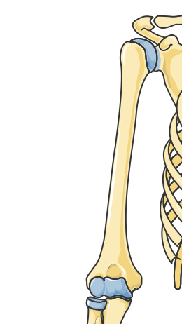
Bones of the Upper Limb
The upper limb is highly mobile and versatile, allowing for complex movements and manipulation of objects. This region consists of several bones that work in unison to provide strength and flexibility.
Clavicle
The clavicle, also known as the collar bone, serves as a connection between the arm and the trunk, acting as a support strut that maintains the shoulder in place. It connects the scapula to the sternum, providing stability to the shoulder region. Due to its exposed location, the clavicle is one of the most commonly fractured bones in the body.
Scapula (Shoulder Blade)
Located on the posterior side of the ribcage, the scapula is a flat, triangular bone that serves as the anchor point for many muscles that control shoulder and arm movements, including the rotator cuff muscles. The scapula allows for a wide range of motion in the arm and shoulder.
Humerus
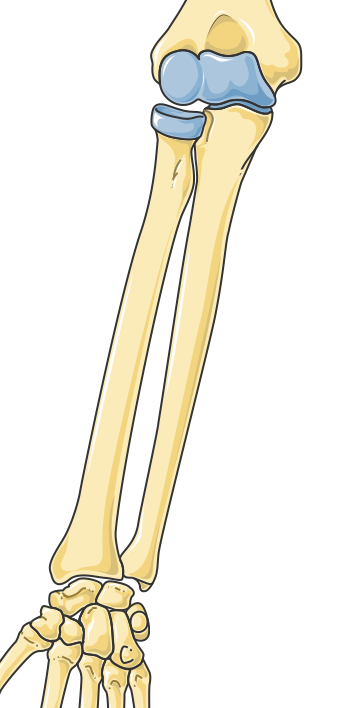
The humerus is the long bone of the upper arm, connecting the shoulder to the elbow. It plays a critical role in arm mobility and provides attachment points for the biceps and triceps muscles. The humerus allows for movements such as flexion, extension, and rotation of the arm.
Radius and Ulna
These two long bones form the forearm.
The radius, located on the lateral (thumb) side, rotates around the ulna to allow for movements such as pronation and supination (rotation of the forearm).
The ulna, positioned on the medial (pinky) side, forms a stable connection with the humerus at the elbow joint, allowing for flexion and extension of the arm.
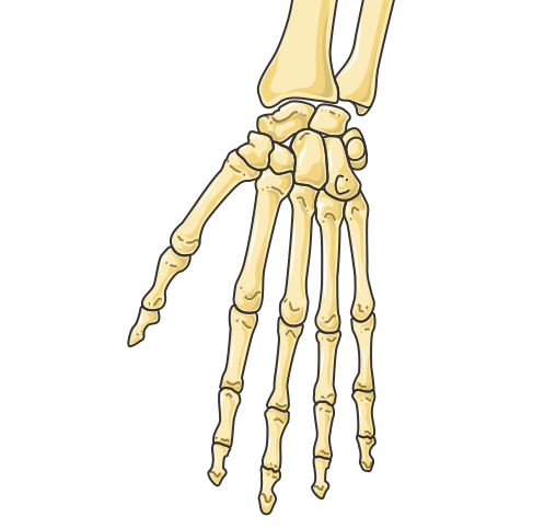
Bones of the Hand
The hand is composed of 27 bones, which provide fine motor control and dexterity. The carpals (wrist bones) connect the hand to the forearm. The metacarpals form the structure of the palm, while the phalanges (finger bones) enable various movements. These bones allow humans to grasp, hold, and manipulate objects with precision.
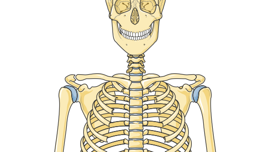
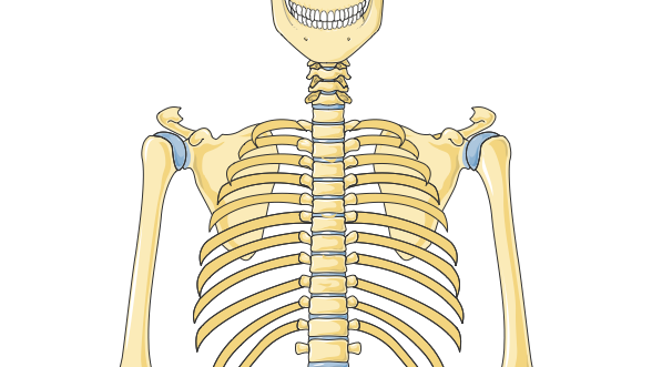
Bones of the Thorax and Spine
The thoracic cage and spine are essential for protecting the body’s vital organs, supporting body weight, and allowing for flexibility and movement.
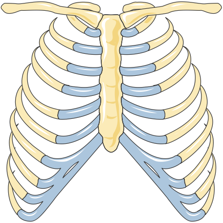
Sternum (Breastbone)
The sternum is a flat bone located in the center of the chest, where it forms part of the ribcage. It anchors the ribs and provides protection for the heart, lungs, and major blood vessels. The sternum consists of three parts:
- Manubrium
- Body
- Xiphoid process.
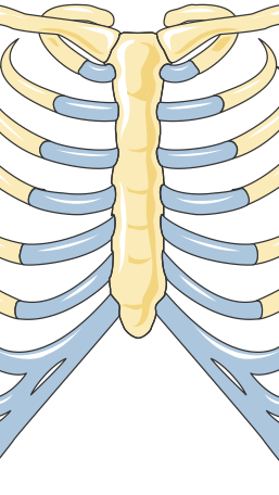
Ribs
The ribcage consists of 12 pairs of ribs that protect the organs within the thoracic cavity, including the lungs and heart. The first seven pairs are known as “true ribs” because they connect directly to the sternum. The next three pairs are “false ribs,” which connect indirectly, and the final two pairs are “floating ribs,” which do not attach to the sternum. The ribs also play a key role in respiration by expanding and contracting the chest during breathing.
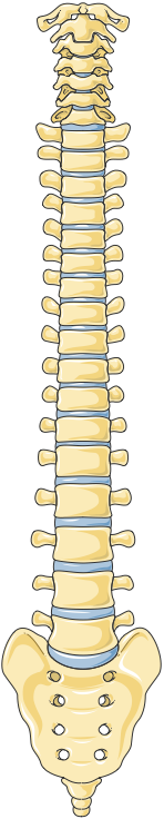
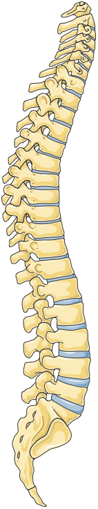
Vertebra
The vertebral column, or spine, is made up of 33 vertebrae, divided into five regions: cervical, thoracic, lumbar, sacral, and coccygeal.
The cervical vertebrae (7) support the head and allow for neck movement.
The thoracic vertebrae (12) anchor the ribs, providing stability to the chest.
The lumbar vertebrae (5) bear the majority of the body’s weight and are crucial for lower back movement.
The sacrum and coccyx are fused vertebrae at the base of the spine, providing a stable foundation for the pelvis and connecting the spine to the lower body.
Bones of the Lower Limb
The bones of the lower limb are designed for weight-bearing, stability, and locomotion. They are among the strongest in the body, supporting activities such as walking, running, and jumping.
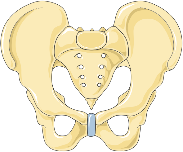
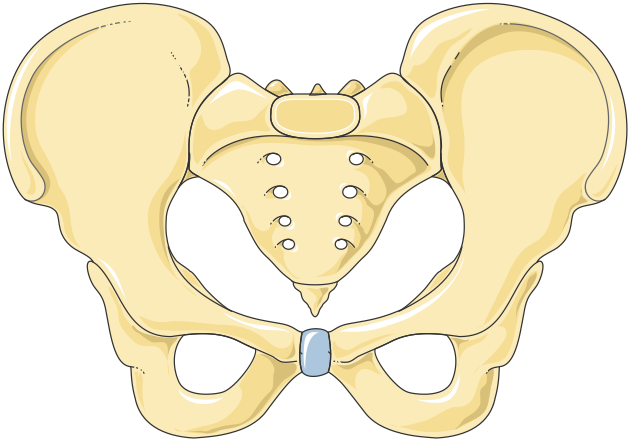
Pelvis
The pelvis is a large, ring-shaped bone composed of the ilium, ischium, and pubis. It serves as the foundation for the spine and connects the trunk to the lower limbs. The pelvis supports the weight of the upper body, protects internal organs such as the bladder and reproductive organs, and provides attachment points for muscles of the lower body.
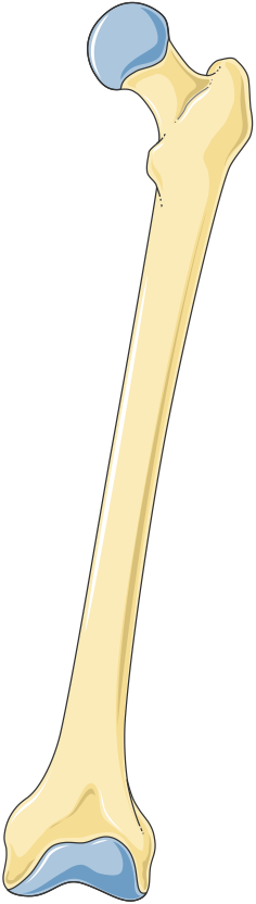
Femur
The femur, or thigh bone, is the longest and strongest bone in the body. It connects the pelvis to the knee and plays a critical role in supporting body weight during movement. The femur is essential for walking, running, and maintaining upright posture, and it provides attachment points for powerful muscles like the quadriceps and hamstrings.
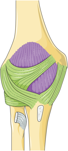
Patella
The patella, or kneecap is a small, flat bone located in front of the knee joint. It protects the knee and improves the leverage of the quadriceps muscle during leg extension, allowing for more efficient movement of the lower leg.
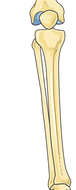
Tibia and Fibula
The tibia, also known as the shinbone, is the larger and stronger of the two lower leg bones. It bears the majority of the body’s weight and connects the knee to the ankle.
The fibula, located parallel to the tibia, provides additional support and stability, particularly for the ankle joint.
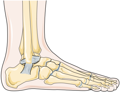
Bones of the Foot
The foot contains 26 bones, which provide support, balance, and mobility. The tarsal bones form the back of the foot and include the heel bone (calcaneus). The metatarsals connect the tarsals to the phalanges, which form the toes. The bones of the foot work together to absorb shock and distribute weight during walking and running, ensuring stability and flexibility.
Common Congenital Anomalies
Congenital anomalies in the skeletal system include structural abnormalities in bones and joints that develop during fetal life. These conditions can range from mild to severe, affecting physical growth, mobility, and overall skeletal function. Early diagnosis and management are essential, particularly for conditions that may impair motor abilities or cause deformities. Here are some of the most common congenital skeletal anomalies:
| Congenital Skeletal Anomaly | Description |
|---|---|
| Clubfoot | Congenital Talipes Equinovarus (Clubfoot )is a deformity where one or both feet are twisted inward and downward. It affects about 1 in 1,000 newborns and requires early intervention. Treatment often involves a combination of stretching, casting (Ponseti method), and, in some cases, surgical correction to improve foot alignment and function. |
| Developmental Dysplasia of the Hip (DDH) | DDH is a condition where the hip joint is improperly formed, allowing the femoral head to dislocate from the acetabulum. It is often detected in infancy. Treatment includes bracing with a Pavlik harness in mild cases or surgical intervention in more severe cases to stabilize the joint and promote normal hip development. |
| Achondroplasia | Achondroplasia is the most common form of dwarfism, caused by abnormal cartilage formation leading to shorter limb bones. Individuals have a characteristic body shape with shortened arms and legs, while the torso remains relatively normal in size. Management focuses on supportive care, including physical therapy, and addressing potential complications like spinal stenosis. |
| Osteogenesis Imperfecta (OI) | Also known as “brittle bone disease,” OI is a genetic condition that results in fragile bones prone to fractures, often from minor trauma. Treatment includes fracture management, physical therapy, and in some cases, medications like bisphosphonates to strengthen bones. |
| Spinal Muscular Atrophy (SMA) | Although primarily a neuromuscular disorder, SMA often results in secondary skeletal issues due to muscle weakness and spine curvature. Progressive weakness can lead to scoliosis and joint contractures. Management includes physical therapy, orthopedic support, and, in severe cases, surgical intervention for scoliosis. |
| Syndactyly and Polydactyly | Syndactyly refers to the fusion of fingers or toes, while polydactyly involves extra fingers or toes. Both conditions can be surgically corrected, often in early childhood, to improve hand or foot functionality and appearance. |
| Craniosynostosis | Craniosynostosis is the premature fusion of one or more cranial sutures, which can affect skull shape and restrict brain growth. Symptoms may include an abnormal head shape and increased intracranial pressure. Surgical intervention is usually required to correct skull shape and allow normal brain development. |