Anatomia układu płciowego męskiego | Male Reproductive System Anatomy
Did You Know?
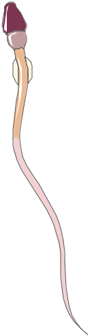
Sperm must navigate a journey from the cervix, through the uterus, and into the fallopian tubes—a distance of about 15 to 18 centimeters. To put this in perspective, it’s like a human swimming the length of an Olympic-sized pool 100 times!
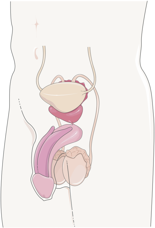
Structure of the Male Reproductive System
The male reproductive system is a complex network of organs designed for the production, maturation, and delivery of sperm. It is divided into two primary components:
- External Genitalia: Includes the penis and scrotum, which are key for sexual function and reproduction.
- Internal Genitalia: Comprises the testes, epididymis, vas deferens, seminal vesicles, prostate gland, and bulbourethral glands, all essential for sperm production and transport.
Male External Genital Organs
The Penis
The penis is the male organ involved in both sexual intercourse and urination. It consists of three columns of erectile tissue: two corpora cavernosa on the dorsal side and one corpus spongiosum on the ventral side, which surrounds the urethra. During sexual arousal, these tissues fill with blood, resulting in an erection. The process is regulated by the parasympathetic nervous system, while ejaculation is controlled by the sympathetic nervous system.
- Glans Penis: The sensitive, rounded tip of the penis, crucial for sexual stimulation. It is densely packed with nerve endings that enhance the sensation during intercourse.
- Prepuce (Foreskin): A retractable fold of skin that covers the glans, providing protection and lubrication. It can be removed through circumcision, a procedure often performed for cultural, religious, or medical reasons.
- Urethra: The urethra runs through the corpus spongiosum, serving as a passage for both urine and semen. It extends from the bladder, through the prostate, and along the length of the penis.
The Scrotum
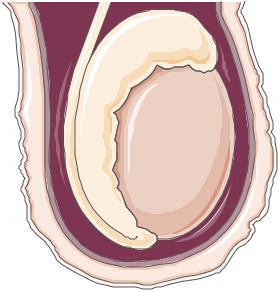
The scrotum is a sac-like structure that supports and protects the testes. It plays a crucial role in maintaining an optimal temperature for spermatogenesis, which is typically 2-3°C lower than body temperature. This is vital for effective sperm production and survival.
- Dartos Muscle: A layer of smooth muscle in the scrotal wall that wrinkles the skin to reduce surface area, thereby conserving heat. It helps in maintaining the temperature of the testes, crucial for spermatogenesis.
- Cremaster Muscle: A muscle that raises and lowers the testes to regulate their temperature, keeping them slightly cooler than the rest of the body. This muscle contracts in response to cold temperatures or physical touch, bringing the testes closer to the body for warmth.
Male Internal Genital Organs
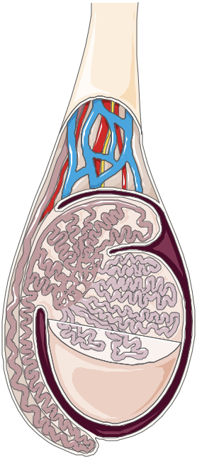
The Testes (Testicles)
The testes are the primary reproductive organs in males, responsible for producing both sperm and testosterone. Each testis is enclosed in a tough, fibrous capsule known as the tunica albuginea, which extends inward to divide the testis into approximately 250 lobules. Each lobule contains coiled seminiferous tubules, where spermatogenesis (sperm production) occurs. Spermatogenesis takes about 64 days and is regulated by the hormones FSH (follicle-stimulating hormone) and LH (luteinizing hormone) from the pituitary gland.
- Seminiferous Tubules: These coiled structures are the site of sperm production. They are lined with Sertoli cells, which support and nourish developing sperm cells.
- Leydig Cells: Located in the connective tissue between the seminiferous tubules, they produce testosterone, the primary male sex hormone. Testosterone is essential for the development of male secondary sexual characteristics, libido, and maintenance of muscle mass and bone density.
The Epididymis
The epididymis is a tightly coiled tube that sits along the posterior surface of each testis. It serves as a storage and maturation site for sperm. Sperm gain motility and the ability to fertilize an egg as they pass through the epididymis, a process that takes about 2 weeks.
- Head: Receives immature sperm from the testes through the efferent ducts.
- Body: The central region where sperm maturation continues. Sperm undergo further biochemical changes here, acquiring the ability to move actively.
- Tail: Stores mature sperm until ejaculation. Sperm can be stored in the tail of the epididymis for several days or even weeks until they are either ejaculated or reabsorbed by the body.
The Vas Deferens (Ductus Deferens)
The vas deferens is a muscular tube that transports mature sperm from the epididymis to the urethra in preparation for ejaculation. It is approximately 30-45 cm long and is lined with a thick layer of smooth muscle, which contracts rhythmically during ejaculation to propel the sperm forward.
- Ampulla: The widened end of the vas deferens that acts as a temporary sperm reservoir. It plays a role in the temporary storage of sperm before ejaculation.
- Spermatic Cord: Contains the vas deferens, testicular artery, veins of the pampiniform plexus, lymphatic vessels, and nerves. It supports the testis and regulates the temperature via the pampiniform plexus, a network of veins that cool the blood before it reaches the testes.
The Seminal Vesicles
The seminal vesicles are paired glands located behind the bladder. They secrete a viscous, fructose-rich fluid that constitutes the majority of the ejaculate volume (approximately 60-70%). This fluid provides energy for sperm and contains prostaglandins, which help in sperm motility and the thinning of cervical mucus.
- Ejaculatory Ducts: Formed by the union of the vas deferens and seminal vesicle ducts, these ducts pass through the prostate and empty their contents into the prostatic urethra. They are critical in the mixing of sperm with seminal fluid.
The Prostate Gland
The prostate gland, about the size of a walnut, surrounds the urethra just below the bladder. It produces a milky, slightly acidic fluid that enhances sperm motility and viability. This fluid, which makes up about 20-30% of the semen volume, contains enzymes (such as PSA—prostate-specific antigen), zinc, and citric acid, which play important roles in sperm health and the liquefaction of semen after ejaculation.
- Prostatic Urethra: The segment of the urethra that runs through the prostate, allowing the prostatic fluid to mix with sperm. This portion is crucial for the seamless passage of semen during ejaculation.
The Bulbourethral Glands (Cowper’s Glands)
These small, pea-sized glands are located below the prostate near the base of the penis. They secrete a clear, mucus-like fluid during sexual arousal, known as pre-ejaculate, which lubricates the urethra and neutralizes any acidic urine residue. This pre-ejaculate fluid also helps to reduce friction during intercourse.
- Ducts of Bulbourethral Glands: Transport the pre-ejaculate fluid into the spongy urethra, ensuring a smooth passage for semen during ejaculation.
Supporting Structures
The Spermatic Cord
The spermatic cord consists of the vas deferens, blood vessels, lymphatics, nerves, and the cremaster muscle. It extends from the inguinal canal to the testes, playing a vital role in the support and temperature regulation of the testes. The pampiniform plexus within the spermatic cord acts as a heat exchanger, cooling the arterial blood before it reaches the testes, which is crucial for maintaining the optimal temperature for spermatogenesis.
- Pampiniform Plexus: A network of veins that cool the arterial blood before it reaches the testes, helping maintain an optimal temperature for spermatogenesis. This mechanism prevents overheating, which could otherwise impair sperm production.
The Inguinal Canal
The inguinal canal is a passage in the lower abdominal wall through which the spermatic cord passes. It is a common site for hernias, where abdominal contents can protrude into the canal, potentially leading to complications such as pain or obstruction.
Blood Supply and Innervation
Blood Supply
- Testicular Arteries: Arise from the abdominal aorta and supply blood to the testes. They travel through the spermatic cord to support testicular function.
- Internal Pudendal Artery: Branches from the internal iliac artery and supplies blood to the penis and scrotum, essential for erectile function.
- Cremasteric Artery: A branch of the inferior epigastric artery that supplies the cremaster muscle, aiding in temperature regulation for the testes.
- Deferential Artery: Originates from the superior vesical artery, supplying the vas deferens to support sperm transport.
Venous Drainage
- Pampiniform Plexus: A network of veins around the testicular artery, aiding in temperature regulation. The right testicular vein drains into the inferior vena cava, and the left drains into the left renal vein.
- Prostatic Venous Plexus: Drains blood from the prostate, connecting with internal iliac veins.
- Penile Venous Drainage: The deep dorsal vein of the penis drains into the internal pudendal veins, supporting venous return post-erection.
Innervation
- Autonomic Innervation: Sympathetic fibers (hypogastric plexus) facilitate ejaculation; parasympathetic fibers (pelvic splanchnic nerves) promote erectile function.
- Pudendal Nerve: Provides sensory and motor innervation to the penis and scrotum, crucial for sensation and ejaculation control.
- Pelvic Splanchnic Nerves: Parasympathetic nerves that innervate the prostate, bladder, and seminal vesicles, aiding in reproductive functions and sexual response.
Common Congenital Anomalies
Congenital anomalies of the male reproductive system involve structural abnormalities of the genital organs that develop during fetal life. These anomalies can range from mild, asymptomatic variations to conditions requiring surgical intervention to prevent complications or preserve fertility. Here are some of the most common congenital anomalies in the male reproductive system:
| Congenital Anomaly | Description |
|---|---|
| Hypospadias | Hypospadias is a condition where the urethral opening is located on the underside of the penis rather than at the tip. This anomaly can affect urination and, in some cases, fertility. Surgery is often performed in early childhood to correct the position of the urethral opening and improve functionality. |
| Cryptorchidism (Undescended Testicle) | Cryptorchidism occurs when one or both testicles fail to descend into the scrotum before birth. This is one of the most common congenital anomalies in male infants, affecting about 3-4% of full-term newborns. Early surgical intervention, typically before the age of one, is recommended to reduce the risk of infertility and testicular cancer. |
| Micropenis | Micropenis is defined as an abnormally small penis due to hormonal or genetic factors. This condition may be associated with other endocrine disorders. Hormonal therapy in infancy or early childhood may stimulate growth, and surgical options may be considered if necessary for function or appearance. |
| Testicular Torsion (Congenital) | While testicular torsion is not exclusively congenital, some individuals have anatomical predispositions (e.g., bell clapper deformity) that increase their risk. Testicular torsion requires emergency intervention to restore blood flow and prevent permanent damage to the testicle. |
| Penile Curvature (Congenital) | Congenital penile curvature, or chordee, occurs when the penis is curved at birth, usually due to differences in tissue structure. While mild curvature may not impact function, more severe cases can lead to difficulties with urination or sexual function. Surgical correction is an option if the curvature affects quality of life. |
| Congenital Absence of the Vas Deferens (CAVD) | CAVD is the absence of one or both vas deferens, the ducts that transport sperm from the testicles. Commonly associated with cystic fibrosis, this condition can lead to infertility. Assisted reproductive technologies, such as sperm retrieval and in vitro fertilization, may be options for affected individuals. |
| Epispadias | Epispadias is a rare condition where the urethral opening is located on the upper side of the penis. It can occur alongside other conditions, such as bladder exstrophy, and may require surgical correction to improve urinary and reproductive function. |